
Hereditary Eye Diseases in Border Collies — A Complete Guide to CEA, PRA, Glaucoma, PLL, and Cataracts
Eye Disease as an Invisible Risk
Border Collies are among the most athletically and mentally demanding dog breeds in existence. They track a frisbee at full sprint, navigate agility courses with split-second precision, and read human body language to anticipate instructions before they’re given. All of this depends on reliable, sharp eyesight.
Yet several hereditary eye diseases have quietly moved through Border Collie lineages for generations. Most are invisible before a clinical event or genetic test reveals them. The diseases differ substantially in timing, genetic mode of inheritance, severity, and clinical urgency — treating them as a single category leads to poor risk management.
This guide covers the five most clinically significant hereditary eye diseases in Border Collies, drawing on peer-reviewed research to explain each condition accurately.
Overview: Five Conditions Compared
| Disease | Gene | Inheritance | Onset | Blindness Risk |
|---|---|---|---|---|
| CEA (Collie Eye Anomaly) | NHEJ1 | Autosomal recessive | Congenital (present at birth) | Low–moderate (severe cases only) |
| PRA-XLPRA3 | RPGR region | X-linked recessive | Progressive from 3–4 years | High (progressive) |
| Primary Glaucoma | OLFML3 | Autosomal recessive | Middle age (sudden onset) | High (emergency surgery required) |
| PLL (Primary Lens Luxation) | ADAMTS17 | Autosomal recessive | 3–8 years | High (emergency surgery required) |
| Hereditary Cataract | HSF4 | Autosomal dominant | 2–7 years | Moderate (progressive) |
CEA (Collie Eye Anomaly) — The Most Prevalent Hereditary Eye Disease
The NHEJ1 Deletion
Collie Eye Anomaly results from a 7.8 kb deletion in intron 4 of the NHEJ1 gene. This variant was identified in 2007 by Parker et al. (Genome Research, 2007) and traces back to a shared ancestor common to several British herding breeds, including Rough and Smooth Collies, Shetland Sheepdogs, and Australian Shepherds.
CEA follows autosomal recessive inheritance: carriers (N/CEA) do not develop the condition, and only dogs homozygous for the deletion (CEA/CEA) are affected.
Prevalence in Border Collies
A large Italian cohort study of 334 Border Collies (Marelli et al., Veterinary Record Open, 2022) found:
- Clear (N/N): 68.9%
- Carrier (N/CEA): 29.6%
- Affected (CEA/CEA): 1.5%
- Mutant allele frequency: 0.163
A carrier rate of approximately 30% is a figure breeders cannot reasonably ignore. By contrast, Rough and Smooth Collies in the United States show affected rates of 80–95%, illustrating that while Border Collies share the same variant, its frequency is substantially lower in the breed (Hitti-Malin et al., Genes, 2023).
Severity Spectrum: Grade 1 to 4
CEA lesions are present at birth and non-progressive — they do not worsen over time. Severity is classified as follows:
| Grade | Finding | Vision Impact |
|---|---|---|
| 1 (mildest) | Choroidal hypoplasia only | Minimal or none |
| 2 | Optic disc coloboma / staphyloma | Mild to moderate |
| 3 | Scleral staphyloma / intraocular hemorrhage | Moderate to severe |
| 4 (most severe) | Retinal detachment | Blindness |
A critical nuance: genotype and phenotype do not correlate reliably. The majority of affected dogs (CEA/CEA) present at Grade 1 or 2 and function normally throughout their lives.
The Diagnostic Window: Weeks 6–8
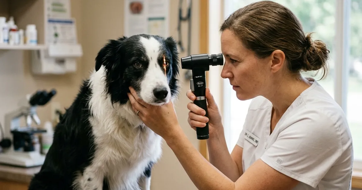
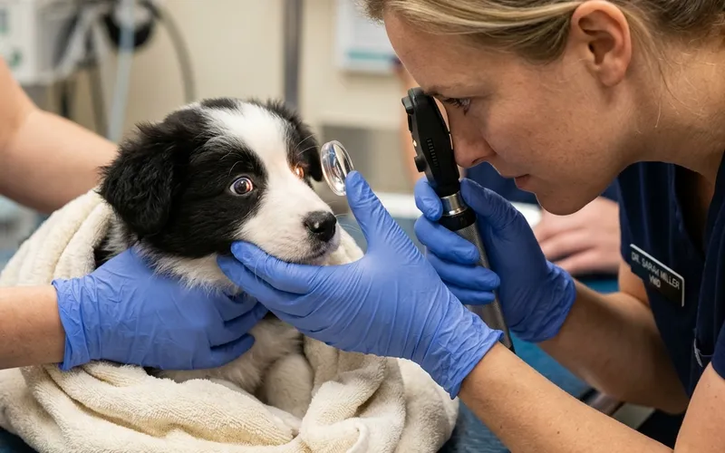
Ophthalmoscopic diagnosis of CEA has a narrow critical window. Between 6 and 8 weeks of age, retinal vasculature is incompletely developed, making choroidal lesions clearly visible. After this period, developing blood vessels can obscure the lesions — a phenomenon called “going normal” — leading to false-negative clinical findings.
The standard practice endorsed by the American College of Veterinary Ophthalmologists (ACVO) is an ophthalmoscopic exam by a board-certified veterinary ophthalmologist between 6 and 8 weeks of age. Genetic testing identifies carrier and affected status but cannot assess phenotypic grade — ophthalmoscopic examination remains essential for that purpose.
PRA (Progressive Retinal Atrophy) — Progressive Blindness in Middle Age
XLPRA3: The Border Collie–Specific Form
The primary PRA affecting Border Collies is XLPRA3, an X-linked recessive form. Because of X-linked inheritance, males need only one copy of the mutation to be affected; females require two copies to be affected, while one copy makes them carriers without clinical disease.
A study in France by Vilboux et al. (BMC Veterinary Research, 2008) examined 487 Border Collies ophthalmoscopically and identified fundic lesions in 60 dogs (12.3%). Of the affected dogs, 54 were male and 6 were female — a sex distribution consistent with X-linked inheritance. The risk allele frequency of r = 0.46 implies a substantial proportion of carrier females in the population.
Disease Progression: Night Blindness to Complete Vision Loss
XLPRA3 progresses in a predictable pattern:
- Ages 3–4: Night blindness begins; difficulty navigating in dim light
- Ages 4–6: Peripheral vision narrows; collision with objects increases
- Age 6+: Daytime vision is affected; complete blindness follows
Electroretinography (ERG) can detect abnormal retinal electrical responses as early as 2 years of age, before clinical signs emerge — enabling pre-symptomatic diagnosis.
Progressive Rod-Cone Degeneration (prcd-PRA), caused by an autosomal recessive mutation in the PRCD gene, is also possible in Border Collies but is reported as very rare or uncommon in North American populations. XLPRA3 represents the dominant clinical concern in the breed.
Primary Glaucoma (Goniodysgenesis) — A Newer, High-Stakes Risk
The Discovery of the OLFML3 Variant
Primary closed-angle glaucoma (PCAG) in Border Collies appears to have emerged in Australia in the late 1990s, subsequently spreading to Europe and North America. In 2019, Oliver et al. published a landmark study in PLOS Genetics (PMC6404605) identifying a missense variant in the OLFML3 gene (c.590G>A; p.Arg197Gln) as the primary cause of goniodysgenesis and subsequent glaucoma in Border Collies.
The variant is inherited as autosomal recessive. The mutant allele frequency in Border Collies was estimated at approximately 5%. When the researchers screened DNA from 12 other dog breeds and wolves, the variant was found only in Border Collies — it appears to be breed-specific.
Genotype-Phenotype Correlation
| Genotype | Glaucoma incidence | Severe Goniodysgenesis |
|---|---|---|
| Homozygous mutant (AA) | 100% (9/9) | 86% (12/14) |
| Heterozygous (AG) | 0% (0/9) | 14% (2/14) |
| Wild type (GG) | 0% (0/9) | 1.5% (1/67) |
The 100% concordance between homozygous status and glaucoma is striking. The authors note incomplete penetrance may exist in some contexts, but homozygosity is a reliable high-risk indicator.
Primary glaucoma causes rapid intraocular pressure elevation, resulting in severe pain, corneal edema, optic nerve damage, and potential permanent blindness within hours. It is a true veterinary emergency.
PLL (Primary Lens Luxation) — Acute-Onset Emergency
ADAMTS17 and the Border Collie Caveat
Primary Lens Luxation occurs when the zonular fibers supporting the lens undergo degeneration, allowing the lens to displace from its normal position. The ADAMTS17 splice-site variant (c.1473+1 G>A) is the established autosomal recessive cause in terrier breeds and was shown to be widespread across multiple breeds (Farias et al., Veterinary Ophthalmology, 2010).
An important caveat applies to Border Collies: the commercially available PLL genetic test targets the ADAMTS17 terrier variant, and the causal variant for PLL in Border Collies has not been identified. A clear result on the ADAMTS17 test does not rule out PLL risk in a Border Collie. Ophthalmoscopic evaluation remains the appropriate screening tool for this breed.
Clinical Urgency
PLL typically presents between 3 and 8 years of age. Anterior lens luxation compresses the iris and cornea, rapidly triggering secondary acute closed-angle glaucoma. This is a time-critical emergency — surgical lens removal within hours of presentation is necessary to preserve vision and control pain.
Signs of acute ocular pain — squinting, eye-rubbing, light sensitivity, a visibly opaque cornea, or a dilated fixed pupil — require immediate evaluation by a veterinary ophthalmologist.
Hereditary Cataract (HSF4) — Dominant Inheritance, One Copy Sufficient
Hereditary cataracts caused by HSF4 (Heat Shock Factor 4) gene variants follow autosomal dominant inheritance — distinguishing them from the other diseases described above. A single copy of the variant is sufficient to increase disease risk, though incomplete penetrance means some carriers remain unaffected.
Onset typically occurs between 2 and 7 years, beginning with posterior subcapsular opacities that can progress to involve the full lens. Surgical phacoemulsification (cataract removal) can restore useful vision, though it involves anesthesia, cost, and postoperative management.
Differentiating hereditary cataracts from age-related nuclear sclerosis or senile cataracts: early onset (under 7 years), bilateral presentation, and a posterior subcapsular pattern are indicators that suggest heritable disease rather than normal aging.
Clear, Carrier, Affected — Understanding the Three Categories
For autosomal recessive diseases (CEA, prcd-PRA, Goniodysgenesis, PLL), genetic test results fall into three categories:
| Result | Genotype | Clinical Impact | Effect on Offspring |
|---|---|---|---|
| Clear | No mutation (N/N) | Does not develop disease | Cannot pass variant |
| Carrier | One copy (N/variant) | Does not develop disease | 50% chance of passing variant |
| Affected | Two copies (variant/variant) | Develops disease | Always passes variant |
A critical point: carriers do not develop disease. A carrier rate of approximately 30% for CEA does not mean 30% of Border Collies have eye disease. Carriers are healthy dogs who carry one copy of the variant and can transmit it to offspring.
Responsible breeding guidelines for autosomal recessive traits:
- Clear × Clear → All offspring clear (ideal)
- Clear × Carrier → 50% clear, 50% carrier; no affected puppies produced
- Carrier × Carrier → 25% affected puppies expected (to be avoided)
- Affected × any → At minimum 50% carriers; affected if bred to carrier or affected
Clear × Carrier is widely accepted by breed health organizations as a valid strategy that prevents affected puppies while preserving genetic diversity.
Note: HSF4 (hereditary cataract) is autosomal dominant, so these rules differ. Even one copy in a parent can pass to 50% of offspring.
When and How to Screen
Genetic Testing: What It Can and Cannot Tell You
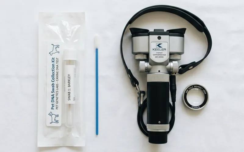
Genetic testing for known variants provides clear, objective pre-breeding data. A swab or blood draw sent to a laboratory can determine whether a dog is clear, carrier, or affected for each tested variant — before any breeding decision is made.
Available platforms include Orivet, Paw Print Genetics, UC Davis Veterinary Genetics Laboratory, and Animal Genetics, among others.
Genetic testing has defined limitations: it detects only known mutations. XLPRA3 does not yet have a commercial genetic test (the causal variant has not been fully characterized). CEA testing identifies genotype but not the severity of clinical lesions. These gaps make ophthalmoscopic examination complementary, not redundant.
Ophthalmologic Examination Schedule
- Weeks 6–8 (puppies): CEA fundus examination by a board-certified ophthalmologist — this diagnostic window lasts only 2–3 weeks
- Annual basis (adults): Retinal screening for PRA, gonioscopy for goniodysgenesis assessment, and lens evaluation for early cataract or subluxation
- Any age (symptomatic): Immediate evaluation for signs of acute glaucoma or lens luxation
The British Veterinary Association and The Kennel Club (UK) operate a joint eye testing scheme (BVA/KC Eye Scheme) for annual screening in predisposed breeds. ACVO/OFA provides a comparable certification program in the United States.
Genetic Disease in Japanese Border Collie Populations
A 2016 study by Mizukami et al. (Veterinary Journal, 2016) assessed the frequency of seven heritable diseases — including CEA, TNS, PRA, and NCL — in more than 100 Border Collies in Japan.
The most striking finding concerned NCL (Neuronal Ceroid Lipofuscinosis, CLN5 variant): in certain Japanese bloodlines, the carrier rate reached 32.9%, with an affected rate of 18.3% and a mutant allele frequency of 34.8%. NCL causes progressive neurological deterioration, visual impairment, behavioral changes, and early death. These figures considerably exceed global frequency estimates.
The authors concluded that genetic counseling combined with systematic breeding management could substantially reduce disease frequency within a few generations.
Prospective puppy buyers in Japan should be aware that untested domestic lines may carry elevated NCL risk, and should request specific NCL (CLN5) test documentation in addition to CEA and PRA screening.
Verifying a Breeder’s Eye Health Screening
Responsible breeders screen breeding dogs before mating and provide documentation. Items to verify:
Genetic testing (minimum recommended panel for eye diseases):
- CEA (NHEJ1)
- Primary Glaucoma / Goniodysgenesis (OLFML3)
- prcd-PRA (PRCD)
- NCL (CLN5) — particularly important in Japan
Ophthalmologic examination:
- Parent dogs: annual eye exam by a board-certified veterinary ophthalmologist (or equivalent)
- Puppies: ophthalmoscopic CEA screening at 6–8 weeks
Questions to ask a breeder:
- “Can you show me the genetic test certificates for CEA, OLFML3, and NCL for both parents?”
- “Were the puppies examined by a veterinary ophthalmologist at 6–8 weeks?”
- “Which laboratory processed the tests, and what are the reference numbers?”
A breeder who states verbally that testing has been performed, but cannot produce certificates from a named laboratory with a specimen ID and test date, has not provided verifiable documentation.
The eyes of a working dog breed are not a secondary consideration. They are the primary means by which a Border Collie navigates the world, reads its environment, and connects with the people it works alongside. Documentation matters: laboratory name, specimen ID, and test date are part of responsible screening.
About ROSCH KENNEL: A Border Collie specialist breeder located at 750 m elevation in Kirishima-Kinkowan National Park, Kagoshima, Japan. All breeding dogs undergo 15+ genetic health tests; results are published in full. Early Neurological Stimulation (ENS) is applied to every litter.
References
- Marelli S. et al., “Genotypic and allelic frequency of a mutation in the NHEJ1 gene associated with collie eye anomaly in dogs in Italy,” Veterinary Record Open, 2022. PMC8800487
- Hitti-Malin RJ. et al., “Global Frequency Analyses of Canine Progressive Rod-Cone Degeneration–Progressive Retinal Atrophy and Collie Eye Anomaly Using Commercial Genetic Testing Data,” Genes, 14(11):2093, 2023.
- Parker HG. et al., “Breed relationships facilitate fine-mapping studies: a 7.8-kilobase deletion cosegregates with Collie eye anomaly across multiple dog breeds,” Genome Research, 17(11):1562–1571, 2007.
- Vilboux T. et al., “Progressive Retinal Atrophy in the Border Collie: A new XLPRA,” BMC Veterinary Research, 4:10, 2008. PMC2324077
- Oliver JAC. et al., “Arginine to Glutamine Variant in Olfactomedin Like 3 (OLFML3) Is a Candidate for Severe Goniodysgenesis and Glaucoma in the Border Collie Dog Breed,” PLOS Genetics, 15(2):e1007975, 2019. PMC6404605
- Farias FHG. et al., “ADAMTS17 mutation associated with primary lens luxation is widespread among breeds,” Veterinary Ophthalmology, 13(6):378–384, 2010.
- Mizukami K. et al., “Molecular prevalence of multiple genetic disorders in Border collies in Japan and recommendations for genetic counselling,” Veterinary Journal, 212:56–61, 2016. PMID:27387721
Daily observation is where the next article begins.
Instagram shows the dogs in daily life. The journal turns the themes we notice there into evidence-aware resources.